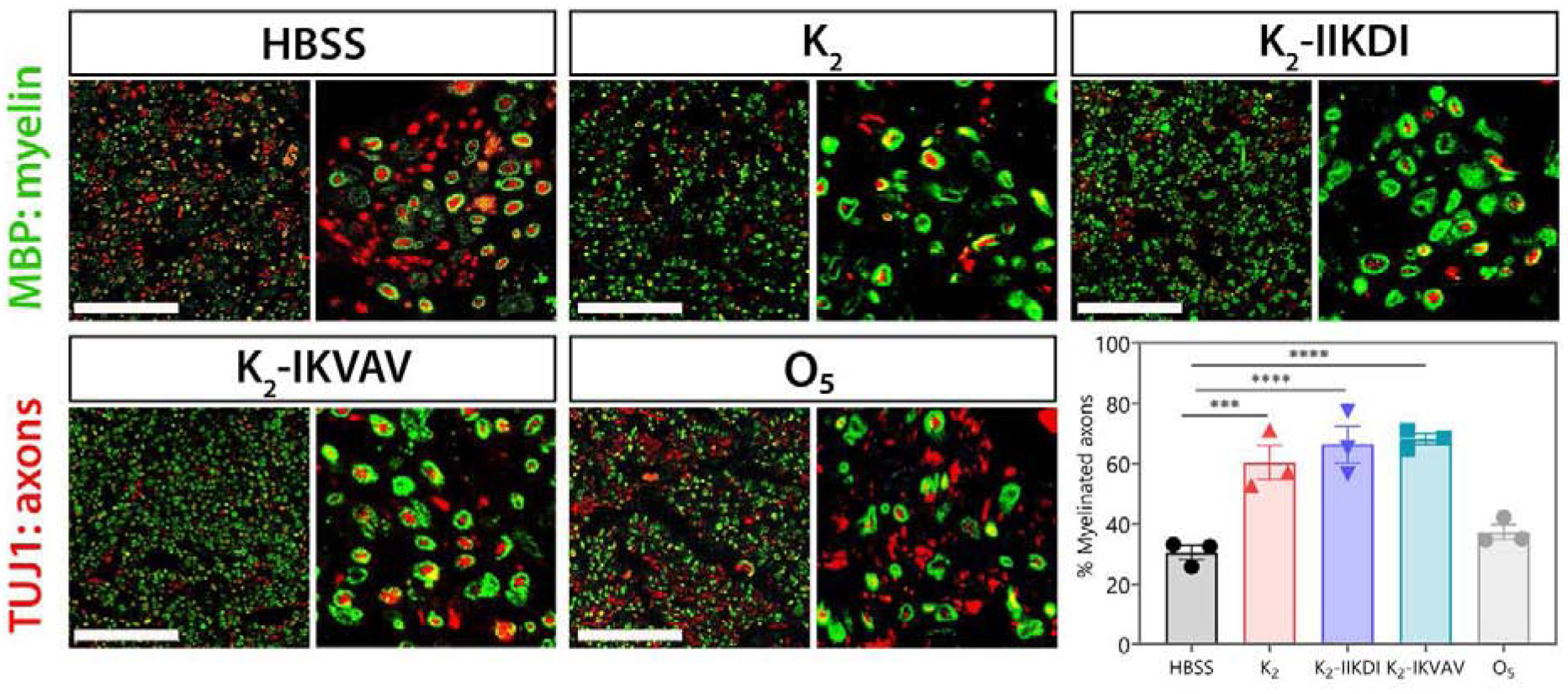
Figure 6.

Sciatic nerves show differential myelination after MDP hydrogel administration. Representative images of sciatic nerve cross-sections stained for TUJ1 (axons) and MBP (myelin) 15 days after crush injury. Cross sections are approximately 800 μm distal from the injury site. Scale bar 100 μm. The fraction of axons that are myelinated are significantly greater in nerves treated with K2, K2-IIKDI, and K2-IKVAV compared to HBSS- or O5-treated nerves. Error bars represent SEM. n = 3 animals per group,. **** p-value <0.0001, *** p-value < 0.0005.
