Figure 7.

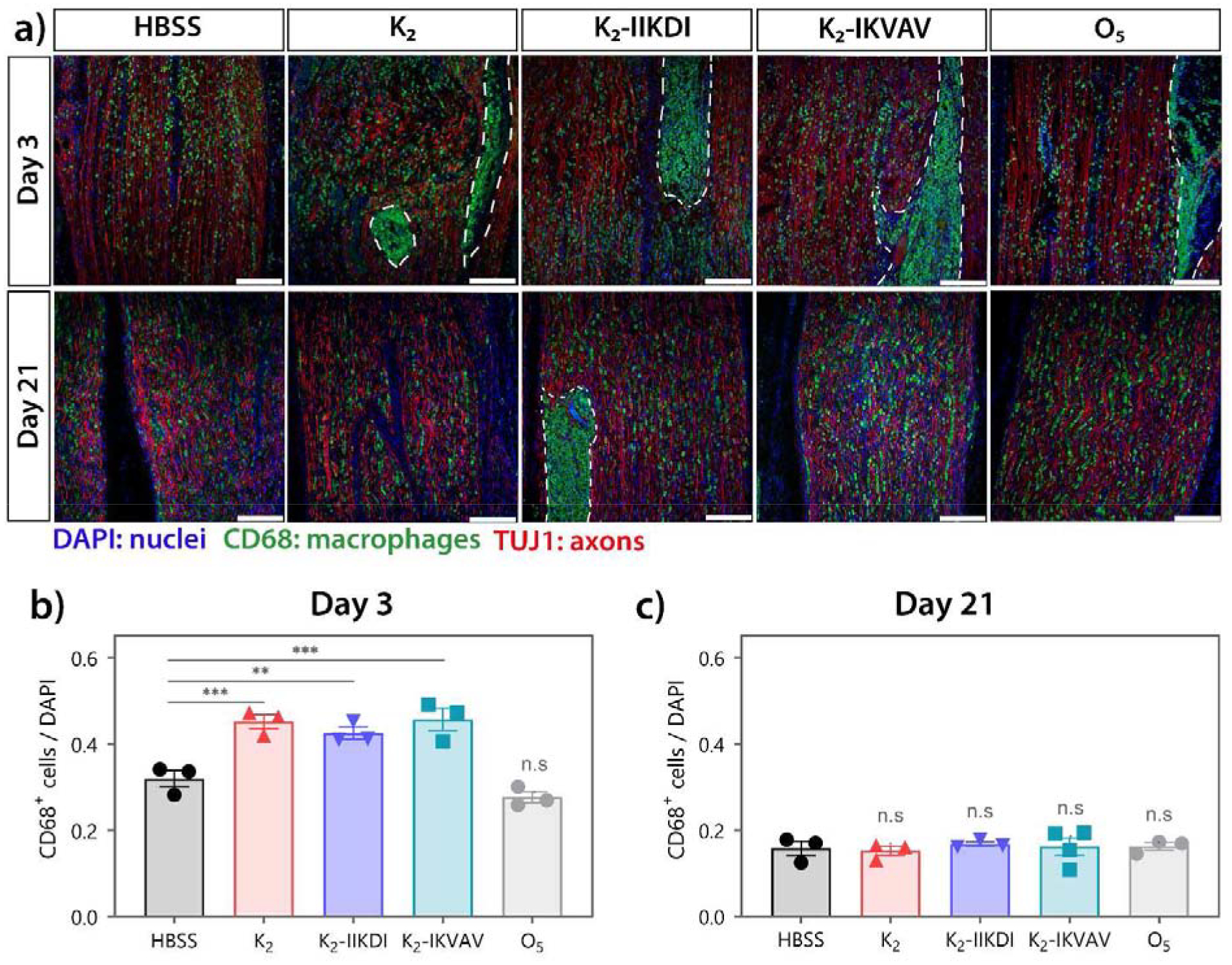
High macrophage infiltration is observed in MDP treated nerves. CD68 and TUJ1 staining of sciatic nerves 3- and 21-days post-injury. a) Representative immunofluorescence images of injured sciatic nerves. Top panel: 3 days post-injury; bottom panel: 21 days post-injury. Dotted line: hydrogel. Scale bar 200 μm. b-c) Quantification of CD68+ macrophages relative to DAPI+ nuclei in areas surrounding hydrogel. Hydrogels were excluded in quantification due to autofluorescence and excessive clustering of cells. b) Nerves treated with K2, K2-IIKDI, and K2-IKVAV had significantly greater CD68+/DAPI+ ratios compared to HBSS-injected control nerves. c) No significant differences were found in CD68+/DAPI ratios between hydrogel-treated and HBSS-injected nerves. Error bars represent SEM. n= 3–4 animals per group. ** p-value < 0.01, *** p-value < 0.001
