Figure 1:
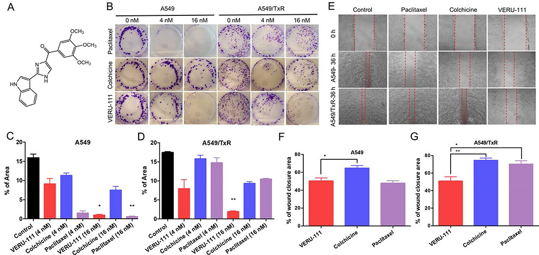
The cell colony formation and migration effects of VERU-111 on A549 and A549/TxR cells. (A) Chemical structure of VERU-111 ((2-(1H-Indol-3-yl)-1H-imidazol-4-yl)(3,4,5-trimethoxyphenyl) Methanone) (B) Representative images from the colony formation assay, (C-D) The quantification of the results from colony area in A549 and A549/TxR cells. The experiment was conducted using two different drug concentrations (4 nM and 16 nM) in a 6-well plate. ImageJ software was used to calculate the total colony occupied area. (E) Would healing assay was done in 12-well plates and the drug solutions were incubated for 36 hours to observe the effects of VERU-111 in compared with positive controls (colchicine and paclitaxel). (F-G) The total scratched area that covered with migrated cells was quantified and represented as percent of total wound closure area. The data is presented as mean ± SEM of triplicates and the Kruskal-Wallis test was performed for the statistical significance. *p < 0.05, **p < 0.01 versus control or indicated.

