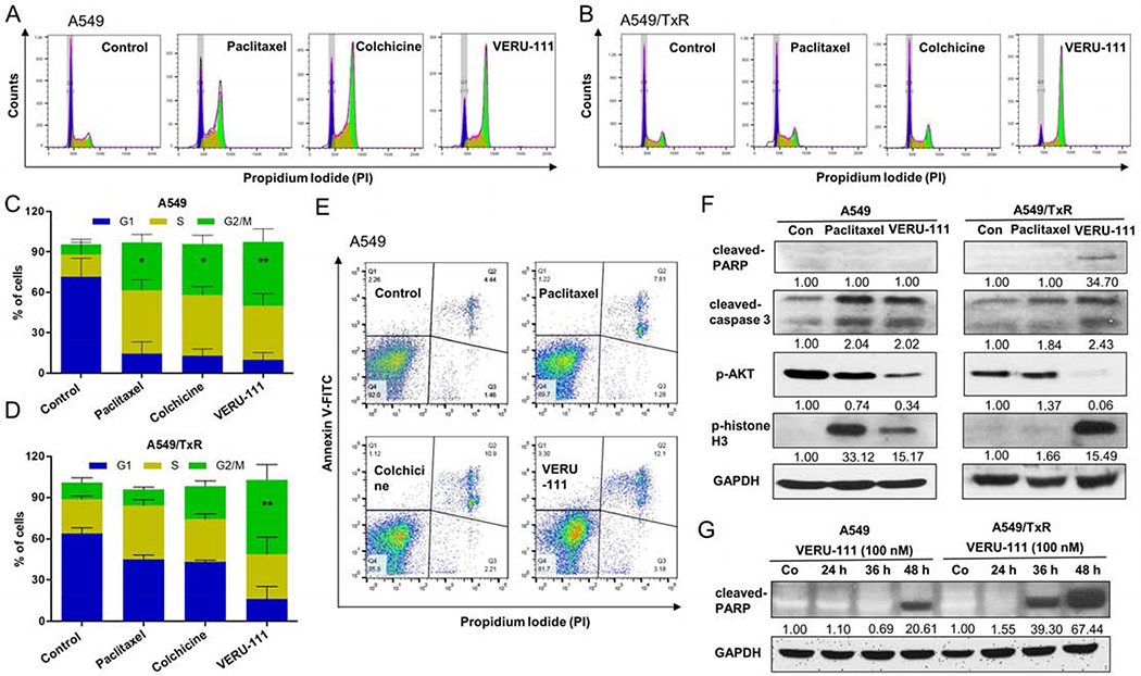
Figure 5:
VERU-111 induces the cell death in A549 and A549/TxR cells. Flow cytometry (FACS) analysis using PI staining showed the cell cycle arrest of G2/M phase by VERU-111 or positive controls in (A) A549 cells or (B) A549/TxR cells after adding 100 nM drug concentration for 24 h. (C-D) The quantitative analysis from figure A and B. The cells were fixed with 70% ethanol and treated with RNase A. (E) VERU-111 displayed highest apoptosis induction in parenteral A549 cells among all groups by annexin V-FITC/PI staining of live single cells. (F) Protein levels of caspase-3 and PARP cleavage, AKT phosphorylation and phosphorylation of histone H3 were identified by western blot assay after treatment with 100 nM paclitaxel or 100 nM VERU-111 for 24 h. GAPDH was used as a loading control. (G) PARP cleavage by western blot following VERU-111 treatment (100 nM) for 24, 36 and 48 h. GAPDH was used as a loading control. Signal intensity was evaluated by Image Studio Lite densitometry, with control group set to 1.00. The data is presented as mean ± SEM of triplicates and Dunnett multiple comparisons were performed for the statistical significance. *p < 0.05, **p < 0.01 versus control or indicated.

