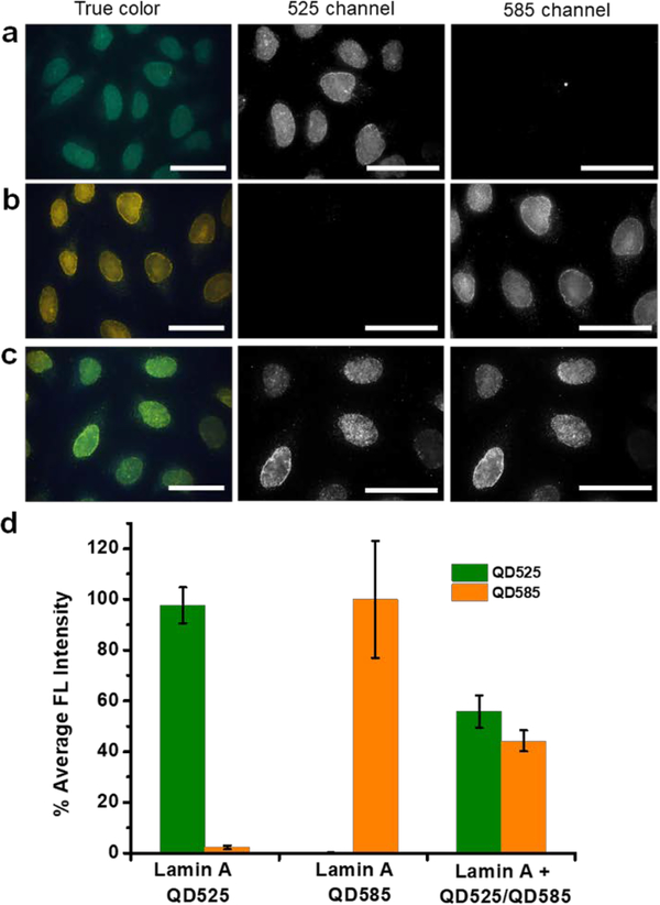
Figure 2. Characterization of QD-SABER potential crosstalk during staining.
a) Pre-assembled QD525-imager mixed with QD585-Streptavidin for Lamin A staining, and b) pre-assembled QD585-imager mixed with QD525-Streptavidin for Lamin A staining. Specific nuclear envelope staining was only observed in the fluorescence channel of the pre-assembled QD-imagers, confirming the absence of imager strands dissociate with the original QD and re-associate with a different QD. c) QD585-streptavidin and QD525-streptavidin of equal molar concentration were mixed with the biotinylated imager probe and applied to cells for Lamin A imaging, signals of similar strength were observed in both fluorescence channels. d) Quantitative bar plots of the cell fluorescence intensity in a), b), and c). HeLa cells were used in this study. Dual-color images were obtained on a microscope equipped with a 100x objective and an HSI camera. Scale bars, 50 μm.

