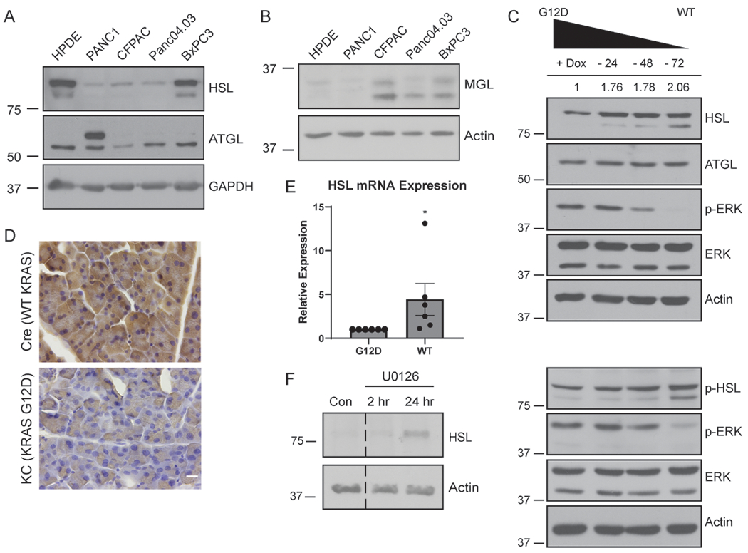
Figure 3.

HSL is regulated by oncogenic KRAS. A, Western blot of the indicated cells for HSL, ATGL, and GAPDH from whole-cell lysates. B, Western blot of the indicated cells for MGL and Actin. C, Western blot of HSL, phospho-HSL, ATGL, phospho-ERK, ERK, and Actin expression from whole-cell lysates. iKRAS cells were cultured in doxycycline, or with doxycycline withdrawn for 24, 48, or 72 hours prior to lysis. Quantitative densitometry values for HSL expression from blot shown (both bands) are indicated above. D, Immunohistochemistry for HSL expression in pancreatic tissue sections isolated from Ptf1a-Cre (Cre) or littermate Ptf1a-Cre driven KRASG12D (KC) mice. n=3 mice per condition. Scale bar, 10 μm. E, Relative HSL mRNA levels by quantitative RT-PCR from iKRAS cells cultured with doxycycline (G12D) or following 72 hour doxycycline withdrawal (WT). 3 technical replicates for 6 independent biological replicates shown. Data analyzed by Wilcoxon Signed Rank test. F, Western blot of mKPC cells treated with vehicle control (Con) or the MEK inhibitor U0126 for the indicated time. Blots are representative of three independent experiments. Graphs indicate mean ± SEM. *p < 0.05.
