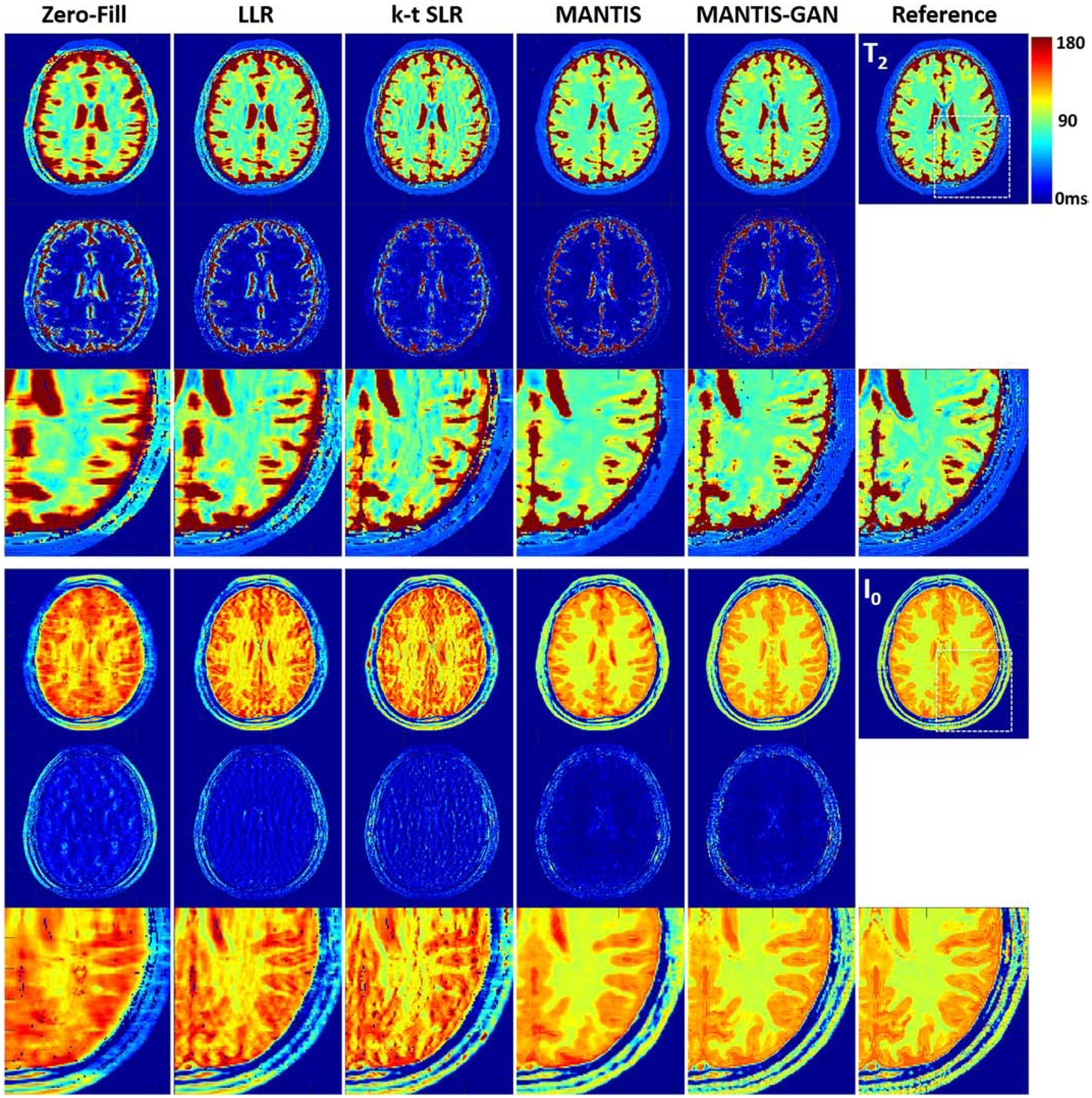
Figure 5:

Comparison of T2 and I0 maps reconstructed from MANTIS-GAN and MANTIS with maps from joint x-p reconstruction methods at an acceleration rate R=8 in one simulated axial brain slice. The difference maps under the reconstructed T2 and I0 maps show the absolute pixel-wise error at the same color scale. MANTIS removed most of the image artifact but resulted in image blurring at the tissue boundaries. MANTIS-GAN generated nearly artifact-free T2 and I0 maps with well-preserved image sharpness, clarity, and tissue texture like the reference maps. The joint x-p reconstruction methods resulted in parameter maps at reduced image sharpness and remaining residual artifacts.
