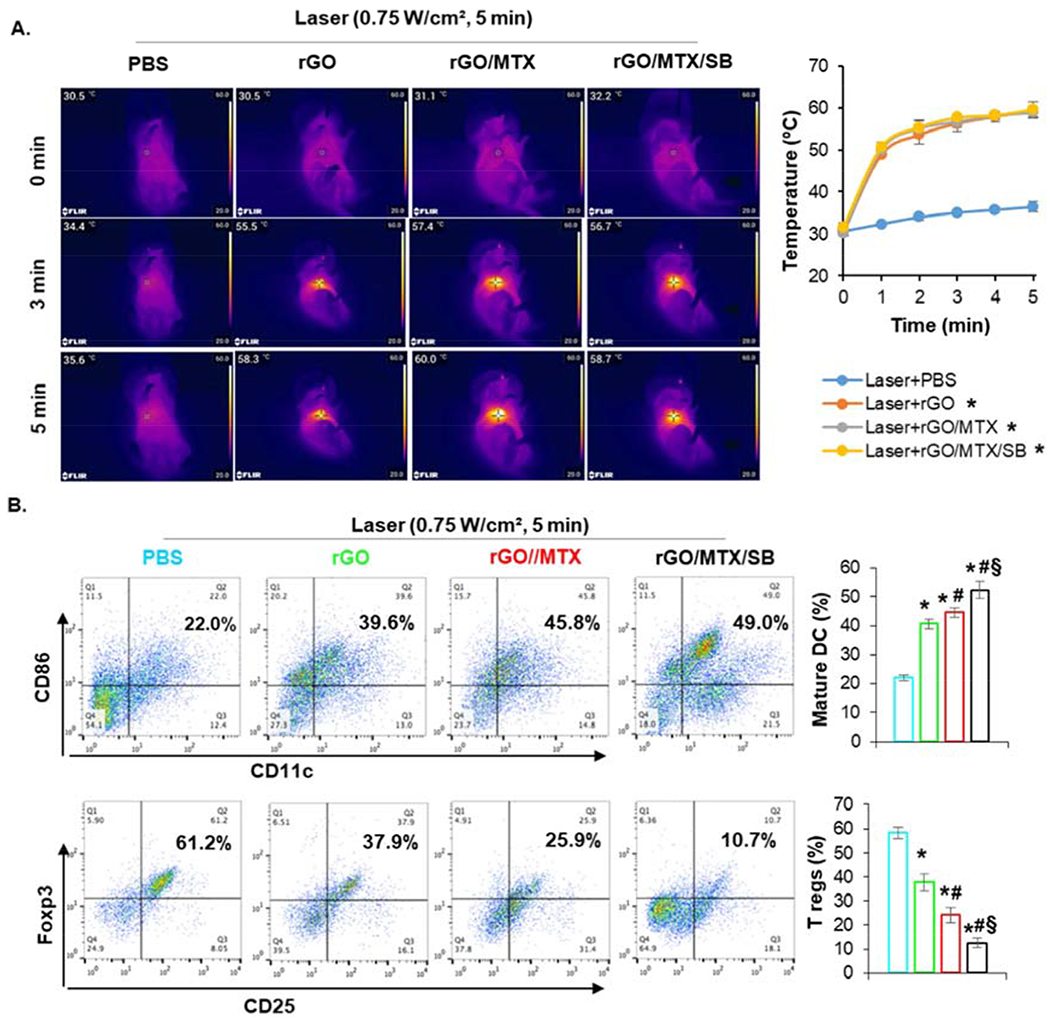
Fig. 5.

Effects of rGO/MTX/SB based PTT on treated primary tumors. A. Temperature increase in tumor tissue under different treatments. Left: IR thermal images of 4T1 tumor bearing mice treated with different rGO derivatives under an 805-nm laser irradiation (0.75 W/cm2 for 5 min). Right: Temperature increase on the surface of tumor tissue during laser irradiation. B. Infiltration of matured DCs and Tregs in treated tumor tissue. Dissociated single cells from tumors 24 h after treatments were stained with anti-CD11c, -CD86, -CD25, and -Foxp3 mAbs, analyzed by flow cytometry. Dot plots show CD86+/CD11c+ (marker of matured DCs) and Foxp3+CD25+ (marker of Tregs) cells, while bar graph shows % CD86+ CD11c+ and Foxp3+CD25+ cells for each treatment group, (n = 4, *p<0.001 vs Laser; #p < 0.05 vs Laser + rGO; §p < 0.005 vs Laser + rGO/MTX).
