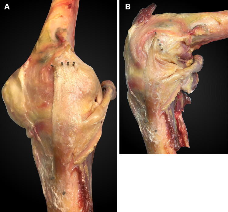
Fig. 1.
Medial aspect of a right knee: a near extension and b at 90° flexion. The four staples in the femur are, from anterior to posterior: the anterior edge of the sMCL, the posterior edge of the sMCL, the anterior edge of the POL, and the posterior edge of the POL. They were hammered completely into the bone after attaching a suture to each of them. The staple loops which guided the anterior and posterior sMCL sutures are visible distally, at the sMCL tibial attachment. The anterior margin of the sMCL wraps around the femoral medial epicondyle with knee flexion. The femoral medial epicondyle is located midway between the anterior and posterior fibre attachments of the sMCL. Note that the distal staples of the POL, in the posterior rim of the tibial plateau, are obscured by the semimembranosus tendon

