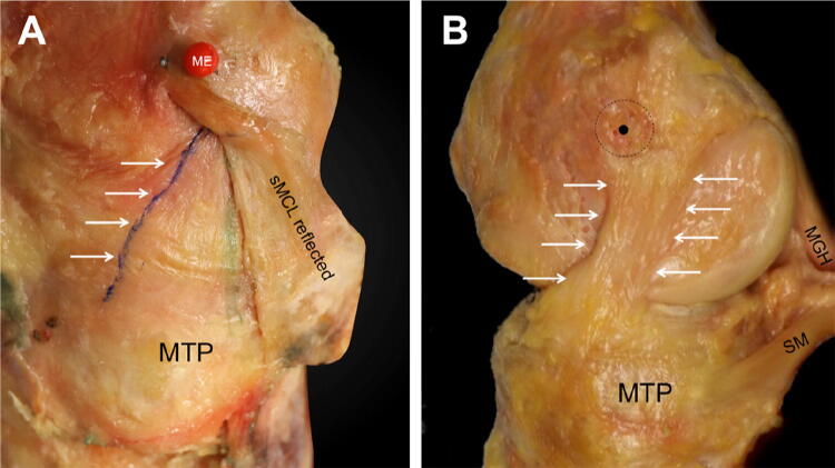
Fig. 2.
a The dMCL of an extended right knee, anterior to the left of the picture and the long axis of the tibia vertically downwards. The red pin head is at the most prominent point of the femoral medial epicondyle (ME): the sMCL attaches anterior and posterior to it. The sMCL femoral attachment is intact, showing the anterior fibres passing anterior to the epicondyle. Distally, the sMCL has been reflected posteriorly (but not released completely) as far as the green line where the sMCL and dMCL blend together to form the PMC, revealing the dMCL, with its most-posterior fibres marked by the green line. The femoral attachment of the dMCL is distal and posterior to the epicondyle, so it is obscured by the sMCL. The most-anterior fibres of the dMCL—the blue line highlighted by the arrows—are oriented anterior/distal from the femur to the tibia. The wrinkle/buckling across the width of the dMCL indicates that it is slack when the sMCL is intact. MTP: medial tibial plateau. b The sMCL has been removed: the most prominent point of the medial epicondyle (black dot) is at the centre of the sMCL attachment (black dashed circle). The oblique antero-distal orientation of the dMCL in neutral tibial rotation is evident, with the anterior and posterior borders indicated by the white arrows. SM direct head of semimembranosus muscle, MGH medial head of gastrocnemius muscle, MTP medial tibia plateau

