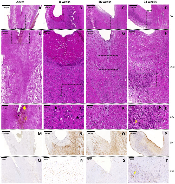
Figure 2.
Hinge region of first-generation TEHVs at different time-points. Representative images of the H&E staining performed on the cross section of the valve. (A–D) 5 × magnification pictures of the hinge region of the valve (scale bars 500 μm). (E–H) 20 × magnification pictures of the hinge region marked by the dotted rectangles in panels (A–D) (scale bars 100 μm). (I–L) 40 × magnification images of the hinge region marked by dotted rectangles in panels (E–H) (scale bars 50 μm). Presence of contractile cells is detected with immunohistochemical images at 8 (N), 16 (O), and 24 (P) weeks. α-SMA+cells are already detected in the hinge area at the earliest time point considered and, over time, these cells are also present in the leaflet. Acute control valve (M) does not present α-SMA+cells. Scale bars: 500 μm, zoom: 5 × . Presence of T-lymphocytes is detected with immunohistochemical images at 8 (R), 16 (S), and 24 (T) weeks. Acute control valve (Q) does not present T-lymphocytes. T-lymphocytes cluster around polymer remnants or in follicles (yellow arrow). Scale bars: 100 μm, zoom: 10 × . In panel I arrow points at leukocytes, star symbol indicates fibrin, and triangle marks erythrocytes. In panel (J) arrow heads point at macrophages and the white arrow indicates eosinophils. In panel (L), the cross symbol shows multinucleated giant cells and the arrowhead macrophages.

