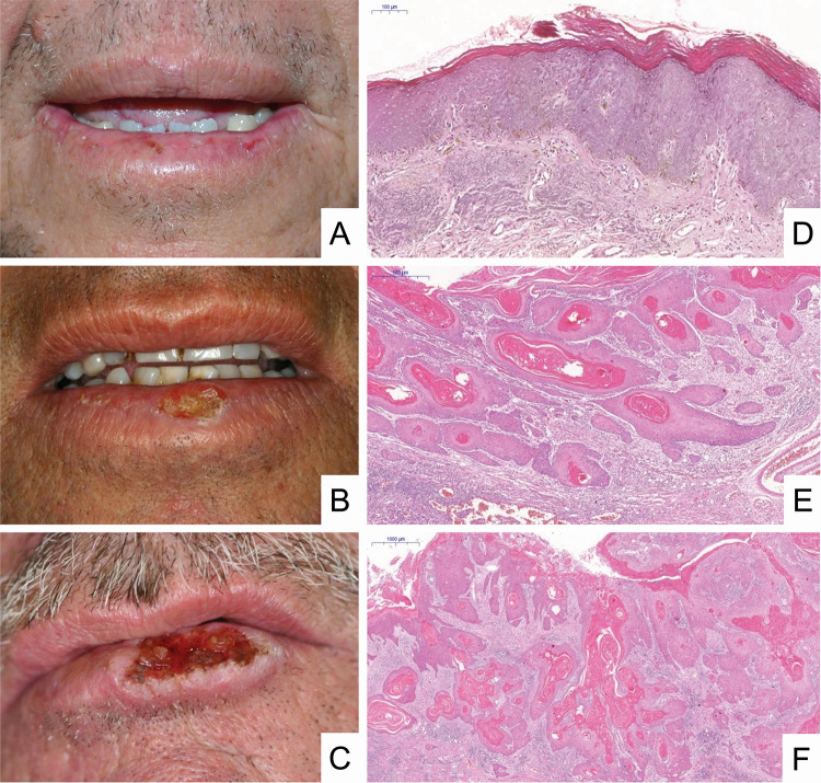
Fig. 3.
Clinical aspects and histopathological features of lip squamous cell carcinoma. a A small ulceration of the lower lip vermilion mimicking actinic cheilitis. Note lesions with crust and ulceration, presence of atrophic regions, oedema, and blurring of the margin between the vermilion and the adjacent skin. b Presence of a crusted lesion with areas of atrophy oedema and blurring of the margin between the vermilion and the adjacent skin. c Presence of an exuberant ulcer with a leucoplastic border, raised and hardened, and beginning of crust formation on the lesion. d In situ carcinoma represented by irregular epithelial stratification, loss of polarity of basal cells, drop-shaped rete ridges and loss of epithelial cell cohesion. In addition, abnormal variation in size and shape is observed in both nucleus and cell, with hyperchromasia, and an increased nucleus/cell ratio [haematoxylin & eosin (H&E), 6 ×]. e Superficially invasive carcinoma represented by nests and cordons of malignant epithelial cells is observed, starting invasion of the lamina propria with formation of keratin pearls and showing an intense inflammatory infiltrate (H&E, 4 ×). f Invasive carcinoma represented by islands of malignant epithelial cells in the lamina propria with intense formation of keratin pearls and an inflammatory infiltrate (H&E, 4 ×).

