
Figure 3.
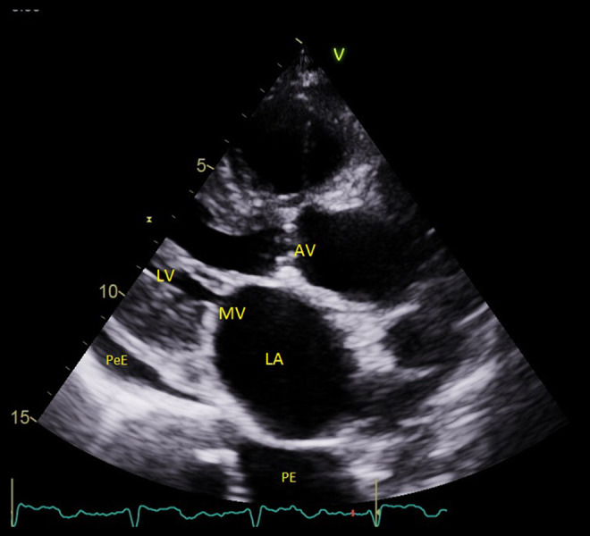
An echo image (parasternal long axis view) from Patient 2. The left ventricle is small, the left atrium is enlarged, the valves are thickened, and there is a small amount of pericardial and plural effusion. AV – aortic valve; LA – left atrium; LV – left ventricle; MV – mitral valve; PeE – pericardial effusion; PE – plural effusion.