
Figure 4.
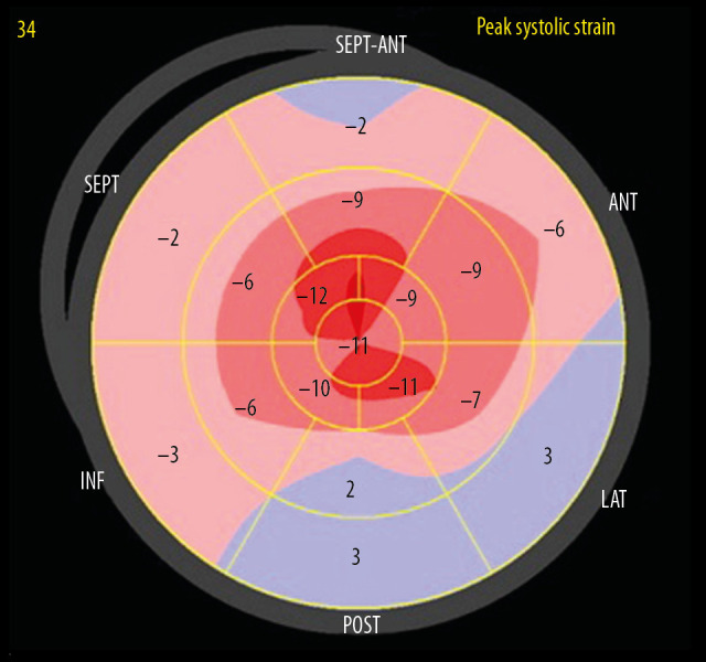
A bullseye display of regional LV longitudinal strain from Patient 2. The basal regions are outermost, mid regions in the middle, and apical regions innermost. A significant reduction in longitudinal strain, with a typical “apical-sparing” or “cherry-on-top” pattern is seen.