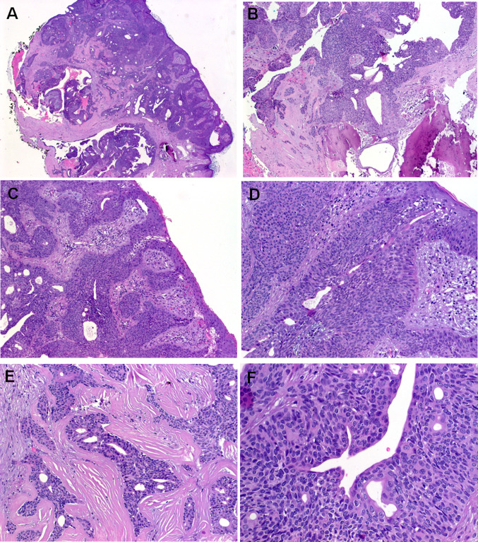
Fig. 3.
Representative images of Case 2. a Whole mount shows basaloid neoplasm with prominent ductal and cystic areas. b Focal erosion of underlying bone is seen. c Prominent replacement of the normal ducts up to the epidermal openings is seen, note lateral spreading replacing the epidermis to variable extent. d Higher magnification of c. e Prominent sclerosis was seen in purely ductal areas. f Solid aggregates of monomorphic basaloid poroid cells are seen surrounding central ducts

