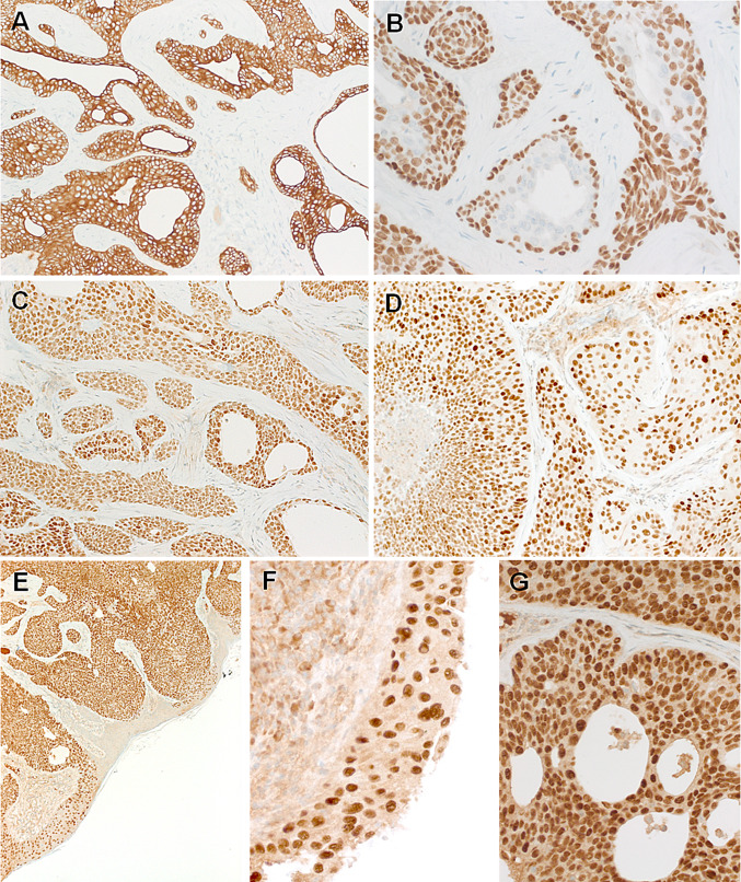
Fig. 4.
Representative images of the immunohistochemistry. Both tumors are strongly positive for CK5 (a) and p63 (b), note focal sparing of ductal cells in the p63 stain (b). SOX10 is expressed in all cells of both tumors (c). Aberrant TP53 is observed in both cases (d). Diffuse NUT expression is seen and it highlights the lateral spreading along the epidermis sparing non-involved epidermal cells (e, f). At high-power, the NUT reactivity is distinctly nuclear (g)

