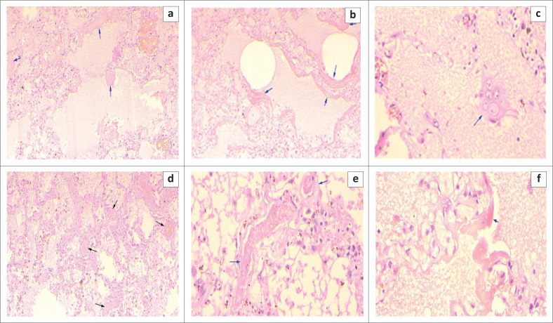
FIGURE 5.
Haematoxylin and eosin staining of lung tissue samples from a 55 year old male patient with COVID-19 (37 Military Hospital, Accra, Ghana, May 2020). Case 3: (a) Severe pulmonary oedema, diffuse alveolar damage with hyaline membrane formation (blue arrow) ×100. (b) Severe pulmonary oedema, diffuse alveolar damage with hyaline membrane formation (blue arrow) ×100. (c) Large pneumocytes (blue arrow) ×400. (d) Microthrombi in small pulmonary arteries (black arrows) ×100. (e) Higher magnification (x400) of image (d) showing microthrombi (blue arrow) ×100. (f) Interstitial widening, pulmonary oedema and prominent hyaline membranes (blue arrow).

