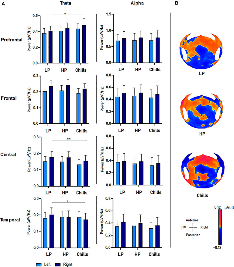
FIGURE 3.
(A) Comparison of power spectral density (μV2/Hz) values in the theta and alpha frequency range for each condition (LP, low pleasure; HP, high pleasure; Chills) in the prefrontal, frontal, central, and temporal ROIs (*p < 0.05, **p < 0.01, errors bars represent SEM). (B) Surface topography for each condition showed an increased positivity for chills and a gradually increasing positivity in parieto-central sites as the emotion increased (μV).

