
Fig. 5.
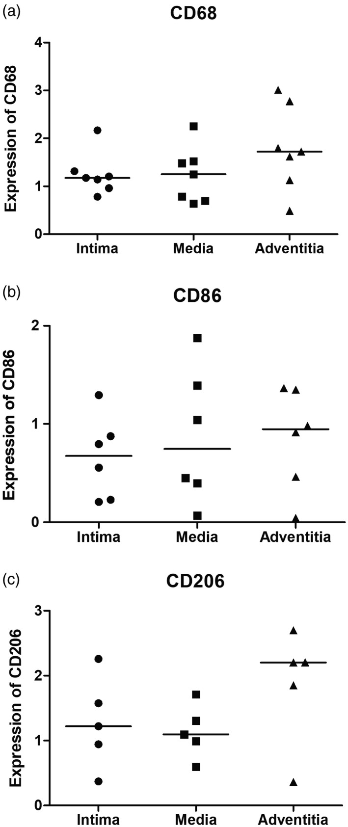
Expression of macrophage markers in the three layers of the aorta from Takayasu arteritis (TAK) patients. No significant differences were observed for the expression of macrophages markers in three layers of the aorta, as follows: CD68 [intima: 1·174 (0·963–1·316) versus media: 1·253 (0·697–1·522) versus adventitia: 1·720 (1·129–2·776); P = 0·328], CD86 [intima: 0·676 (0·223–0·980) versus media: 0·746 (0·316–1·513) versus adventitia: 0·947 (0·358–1·353); P = 0·700] and CD206 [intima: 1·222 (0·658–1·918) versus media: 1·095 (0·793–1·510) versus adventitia: 2·204 (1·107–2·454); P = 0·326]. The crossbar represents the median expression of macrophage markers. Data were presented as median and interquartile range and the analysis was performed by the Kruskal–Wallis test.