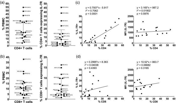
Fig. 4.

CD4+ and CD8+ T cells and their expression of interleukin‐7 receptor (IL‐7R) in acute Kawasaki disease (KD) subjects. The percentages of helper CD4+ T cells (a) and cytotoxic CD8+ T cells (b) in total peripheral blood mononuclear cells (PBMC) are shown as scatter‐plots with median and interquartile range. Subject numbers (Table 1) are shown with the percentage of PBMC in parentheses. We also measured the expression of the IL‐7R on CD4+ and CD8+ populations to explore possible continuous stimulation by antigens. Correlations of the percentage of IL‐7R+ (left panel) or the expression level of IL‐7R [right panel, shown as mean fluorescence intensity (MFI)] with CD4+ (c) and CD8+ T cells (d) are shown.
