
Figure 4.
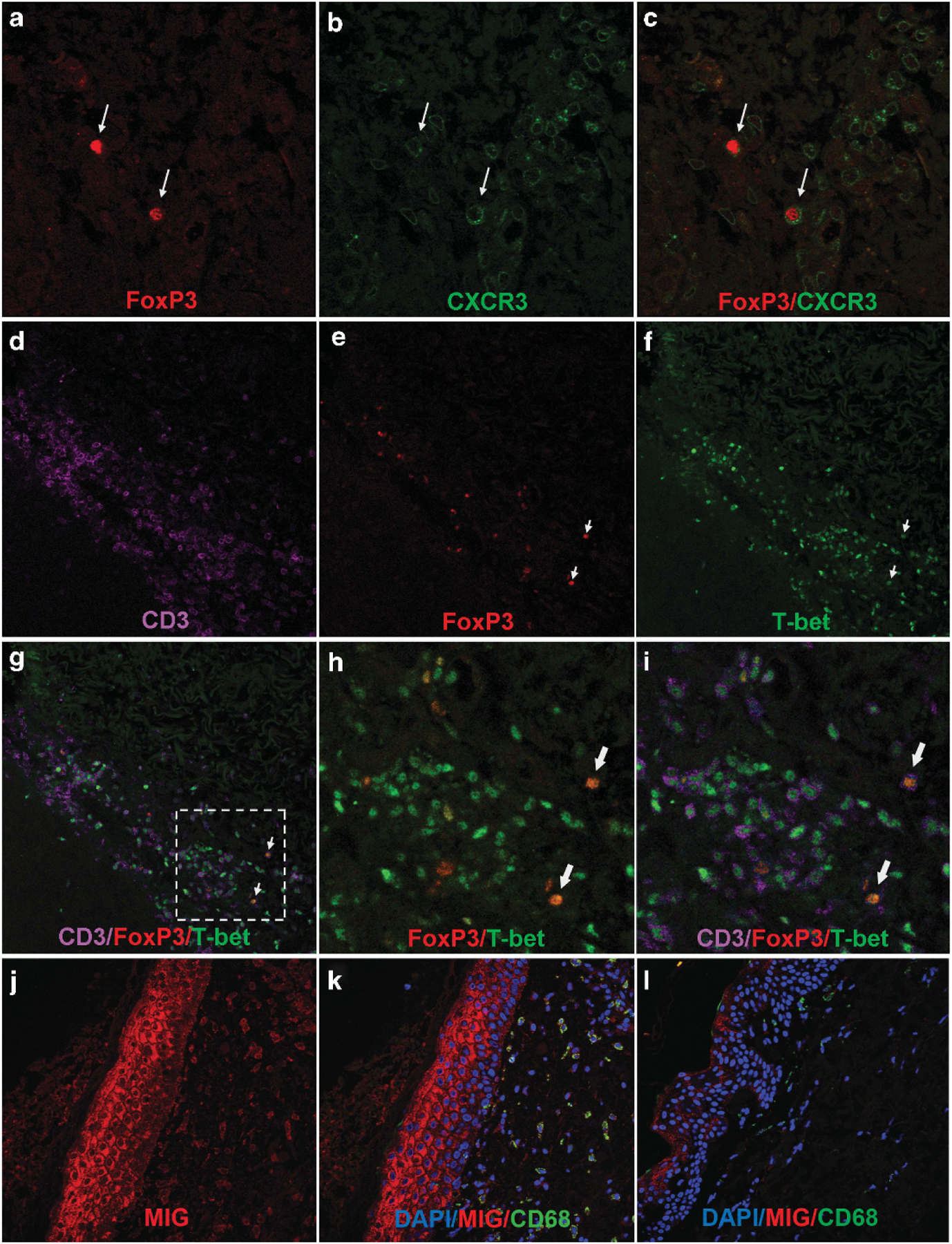
Expression of CXCR3 and T-bet on FoxP3+ T cells in human cGVHD. (a–c) Lichenoid cutaneous cGVHD biopsy stained for FoxP3 (red) and CXCR3 (green). FoxP3+ T cells (arrows) co-express CXCR3. Results typical of seven patients examined. (d–i) Lichenoid cutaneous cGVHD biopsy stained for FoxP3 (red), T-bet (green) and CD3 (lavender). Results typical of seven patients examined. (d–g) Lower magnification showing the dense infiltrate of T-Bet+ CD3+ T cells in lichenoid cGVHD biopsies with scattered FoxP3+ nuclei. (h, i) close-up views of the broken line square in (g) showing colocalization of FoxP3 and T-Bet (combined color orange) (arrows). (j, k) Lichenoid cutaneous cGVHD biopsy (from the tissue block shown in (a–c), stained for MIG (CXCL9) (red), CD68 (green) and DAPI (blue). (j) Both keratinocytes and cells in the dermal infiltrate express MIG (CXCL9). (k) MIG expression co-localizes with CD68, a marker of myeloid cells in the infiltrate. (l) Unaffected skin biopsy collected from a patient with lichenoid cutaneous cGVHD, stained for MIG (CXCL9) (red), CD68 (green) and DAPI (blue). Keratinocytes and the rare infiltrating cells in the dermis express little MIG.