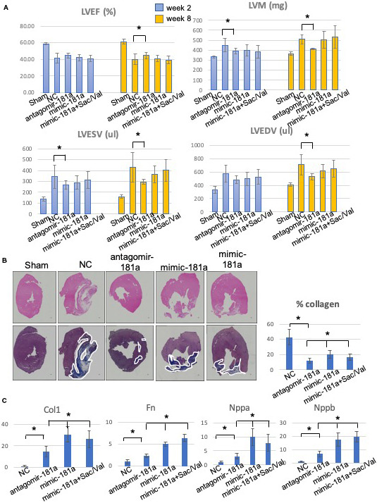
Figure 5. Downregulation of rno‐miR‐181a (mir‐181a antagomir) improves cardiac function and myocardial remodeling processes.

A, Left ventricular ejection fraction (LVEF), left ventricular end‐diastolic volume (LVEDV), left ventricular end systolic volume (LVESV), left ventricular mass (LVM) at weeks 2 and 8. The mir‐181a antagomir treatment group demonstrates significant salutary effects vs the negative control (NC) group. B, Representative images of cardiac tissues in myocardial infarction rats stained with hematoxylin‐eosin (upper panel) and Masson trichrome (lower panel). The myocardial fibers and collagen are colored blue and indicated by white lines. Scale bar=300 μm. Quantitative analysis demonstrated a significant reduction in the percentage of collagen in the miR‐181a antagomir treatment group when compared with NC and the other treatment groups. C, Quantitative polymerase chain reaction analysis for myocardial hypertrophy and fibrosis gene expression in the mir‐181a antagomir treatment group *P<0.05.
