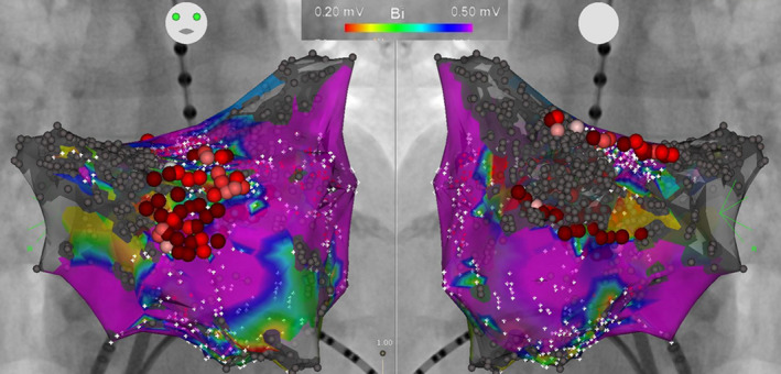
Figure 2. Example of low‐voltage area ablation in addition to pulmonary vein isolation.

Left atrial voltage map after pulmonary vein isolation in a 76‐year‐old female patient. Low‐voltage areas were observed in the anterior‐septal wall and posterior wall. Low‐voltage area ablation consisted of voltage homogenization covering a low‐voltage area in the anterior‐septal wall, and roof and bottom linear ablation isolating a low‐voltage area in the posterior wall.
