
Fig. 5.
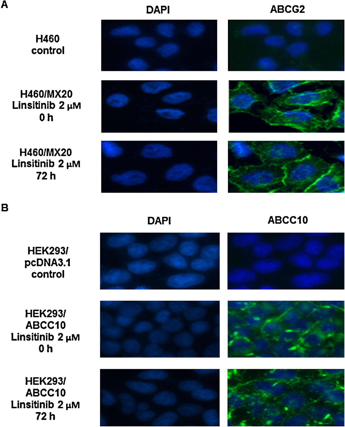
The effect of linsitinib treatment on the subcellular localization of ABCG2 and ABCC10, respectively. (A) H460/MX20 cells were treated with 2 μM linsitinib for 72 h. The subcellular localization of ABCG2 was analyzed by immunofluorescence. ABCG2 staining is shown in green. DAPI (blue) counterstains the nuclei. (B) HEK293/ABCC10 cells were treated with 2 μM linsitinib for 72 h. The subcellular localization of ABCC10 was analyzed by immunofluorescence. ABCC10 staining is shown in green. DAPI (blue) counterstains the nuclei. A representative result was shown and similar results were obtained in two other experiments.