
Figure 4.
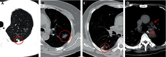
(a) A 46-year-old male COVID-19 patient with fever. CT scan demonstrates a subpleural nodule in the left upper lobe (red circle). (b) A 50-year-old female COVID-19-confirmed patient with dry cough for 4 days. The nontypical halo sign can be seen in the left upper lobe (red circle). (c) The same patient in (a) and the CT displays a nontypical reversed halo sign in the right lower lobe (red circle) in the follow-up CT image. (d) A 60-year-old male COVID-19 patient with initial symptoms of dry cough and fever for 3 days. CT scan shows the enlarged lymph nodes in the mediastinum (red arrows), and this patient is diagnosed coinfection with other bacteria.