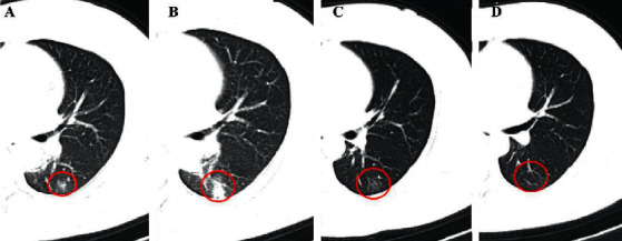
Figure 5.

A 46-year-old female COVID-19 patient. On the initial CT (2 days after the initial symptom onset), the patchy GGO was shown in the left lower lung lobe (a) (red circle), but the lesion progressed to large opacities after approximate 5 days, with more lung tissues involvement (b). After regular treatment in hospital, the majority of the lesion was absorbed and dissipated (14 days), with little linear fibrotic lesions left (c). The lesion was absorbed completely on day 22 (d).
