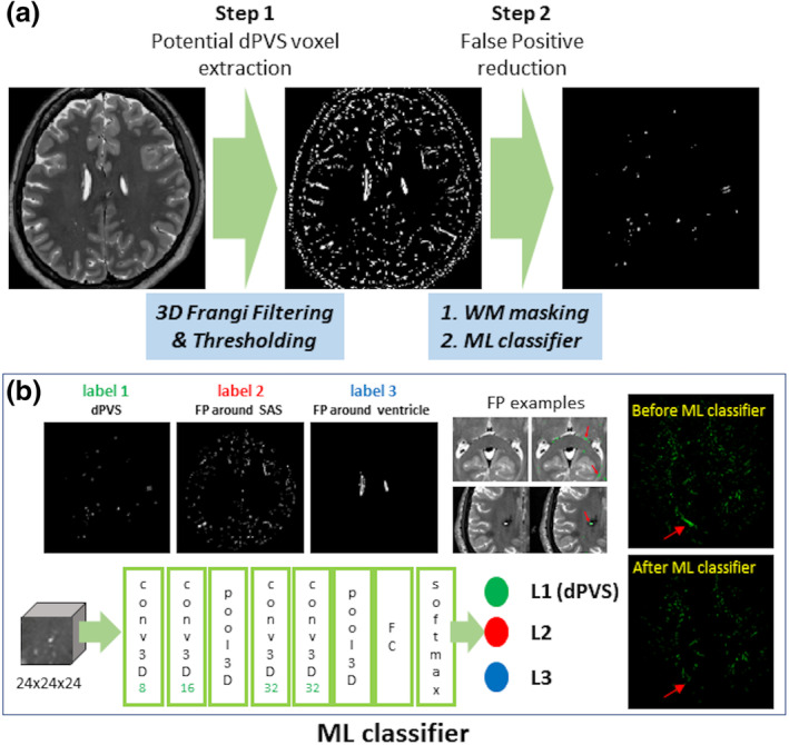
FIGURE 1.

Workflow diagram of fully automated dilated perivascular space (dPVS) segmentation. (a) Extraction of potential dPVS voxels using three‐dimensional Frangi filtering and thresholding with subsequent false‐positive reduction via white matter (WM) masking, and (b) three‐dimensional convolutional neural network (CNN) machine learning classifiers with a 24 × 24 × 24 three‐dimensional input data patch. FP, false positives; ML, machine‐learning; SAS, subarachnoid space
