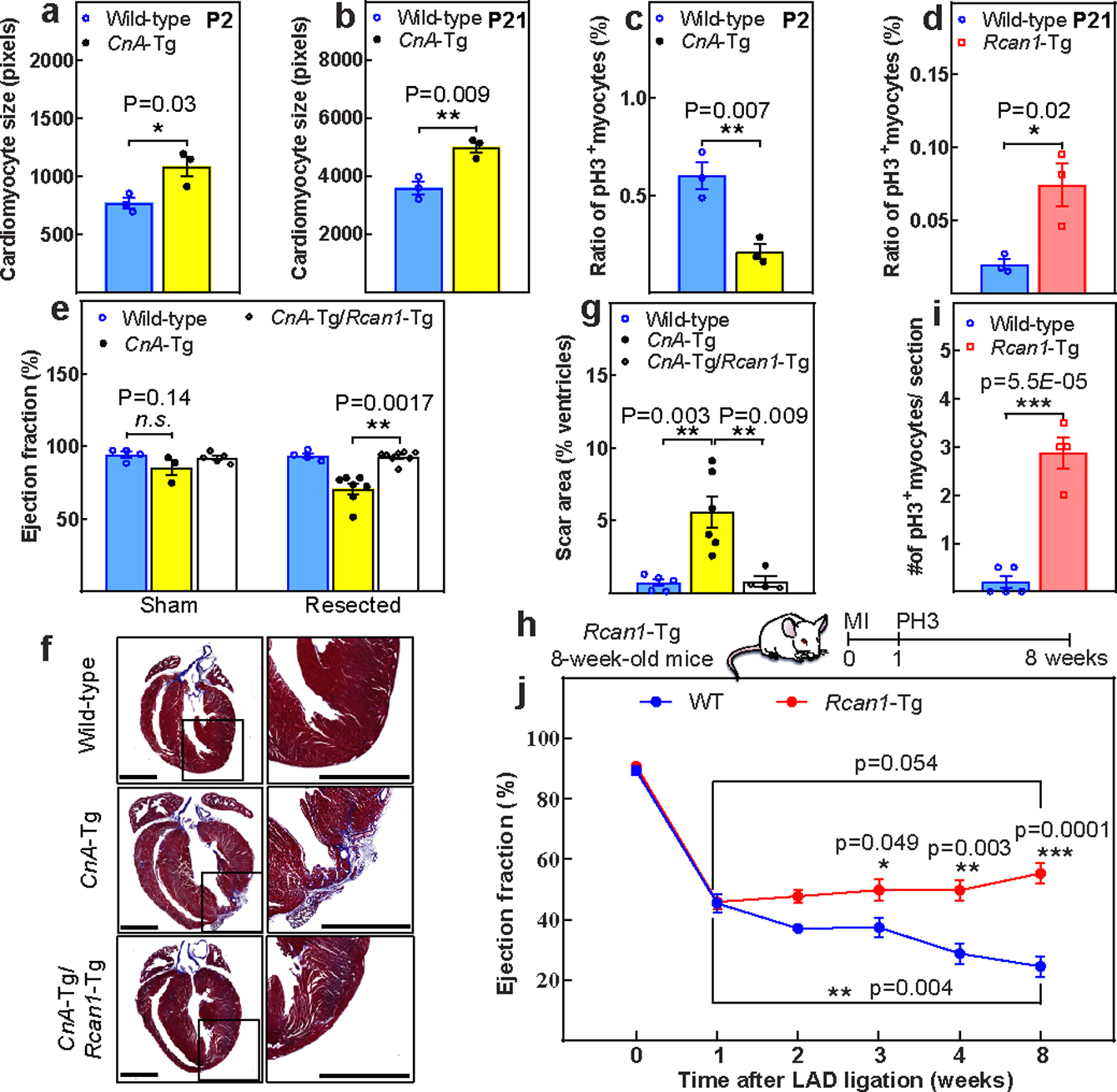
Figure 4: Increased calcineurin activity suppresses postnatal cardiomyocyte proliferation.

a, b, CSA quantification in wild-type and CnA-Tg in P2 (a) and P21 hearts (b). c, d, Percentage of mitotic cardiomyocytes in CnA-Tg P2 (c) or Rcan1-Tg P21 (d) hearts. e, Echocardiographic measurements of cardiac function 21 days after resection. f, g, Representative Masson’s trichrome staining (f; scale bars, 1 mm) and scar area quantification (g) of apical resected hearts 21 days after resection. h, Schematic of MI model in Rcan1-Tg mice. i, Quantification of PH3+ cardiomyocytes in wild-type and Rcan1-Tg heart sections one week after MI induction. j, Serial echocardiographic measurement of wild-type and Rcan1-Tg MI mice. Data are mean ± s.e.m.; unpaired two-sided t-test. *P < 0.05, **P < 0.01, ***P < 0.001. For sample numbers, see Methods. See also Extended Data Fig. 9.
