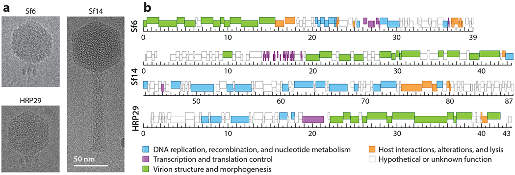
Figure 1.

Characteristics of the developing Shigella phage model systems Sf14 and HRP29 compared with the established model system Sf6. (a) Representative images from cryo-electron micrographs. (b) Genome maps, with genes colored according to function, as indicated. The ruler is in kilo-base pairs. The cluster of short, dark-colored bars at 14–18 kbp in the Sf14 genome represents transfer RNA genes.
