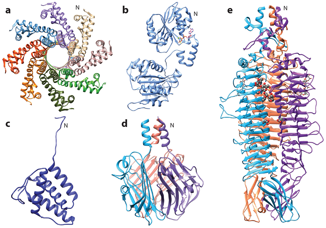
Figure 3.

Structures of Sf6 proteins. (a) Small terminase octamer at 1.65 Å, with the N terminus forming the body facing the viewer and the neck facing away, PDB 3HEF. (b) Large terminase monomer bound to ATPγS at 1.89 Å, PDB 4IEE. (c) Tail adaptor monomer at 1.77 Å, PDB 5VGT. (d) Tail needle knob trimer showing the jellyroll fold at 1 Å, PDB 3RWN. (e) Tailspike trimer with one tetrasaccharide molecule and the catalytic active site residues Asp 399 and Glu 366 shown in gray ball-stick model, resolved to 2 Å, PDB 2VBM. For multimeric proteins, individual subunits are shown in different colors. The N indicates the N terminus for one monomer.
