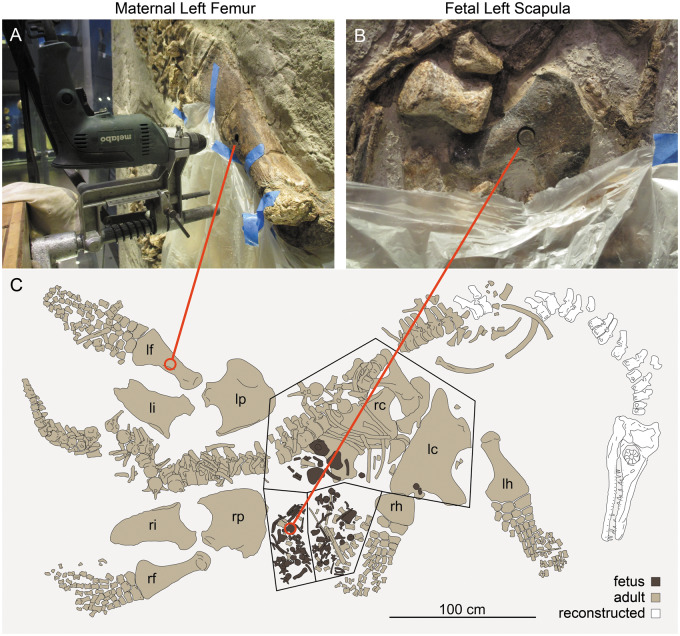
Fig. 1.
Histological sampling of P. latipinnus, LACM 129639. (A) shows jig and core drill in position for sampling of the maternal left femur. (B) depicts sampled core in place within the fetal left scapula; (C) is a schematic of the mount as displayed at the Natural History Museum of Los Angeles County (LACM). lc, left coracoid; lf, left femur; lh, left humerus; li, left ilium; lp, left pubis; rc, right coracoid; rf, right femur; rh, right humerus; ri, right ilium; rp, right pubis.

