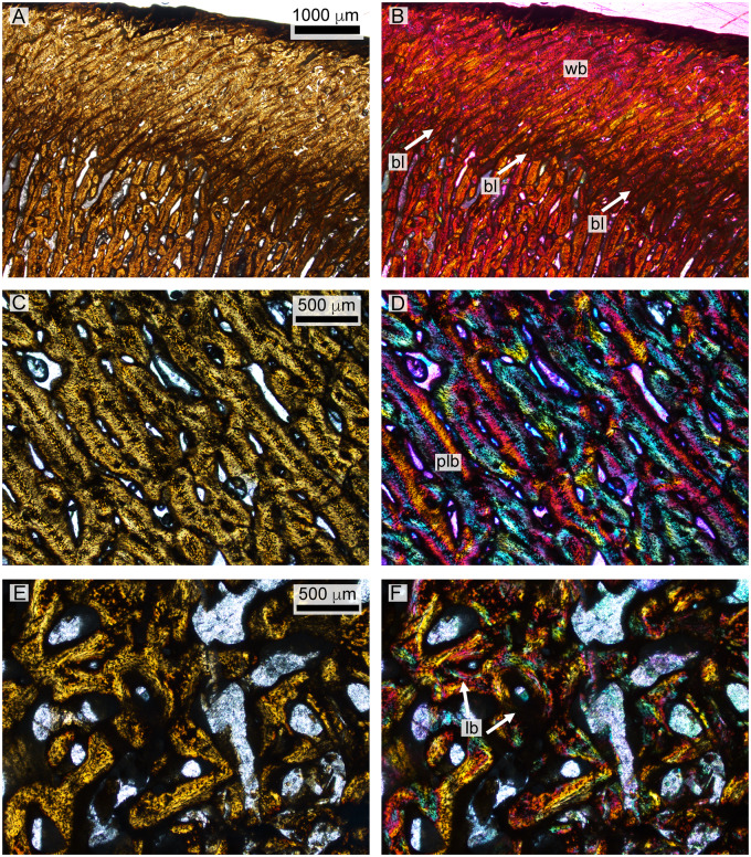
Fig. 12.
Histology of the D. bonneri neonate. (A and B) show the superficial cortex in the upper right quadrant, depicting the birth line in normal and polarized light. (C and D) show the mid-cortical region from the bottom right quadrant in normal and polarized light, consisting of quickly growing radially vascularized fibro-lamellar bone. Panels E and F show vascular cannals and primary lamellar bone from the medullary region in normal and polarized light with lambda filter. bl, birth line; lb, lamellar bone; plb, pseudolamellar bone; wb, woven bone.

