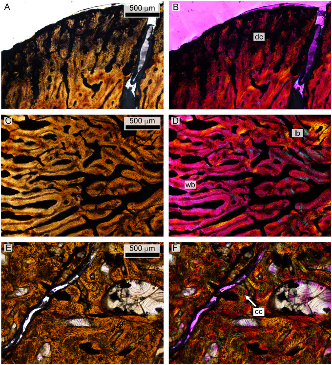
Fig. 13.
Histology of the polycotylid fetus. (A and B) depict the outer edge of the cortex, consisting of poorly ossified woven bone that may be partially decomposed. (C and D) show primary woven bone from the cortex in normal and polarized light with lambda filter. (E and F) illustrate medullary trabeculae composed of primary lamellar bone interspersed with calcified cartilage in normal and polarized light. cc, calcified cartilage; dc, decomposed woven bone; lb, lamellar bone; wb, woven bone.

