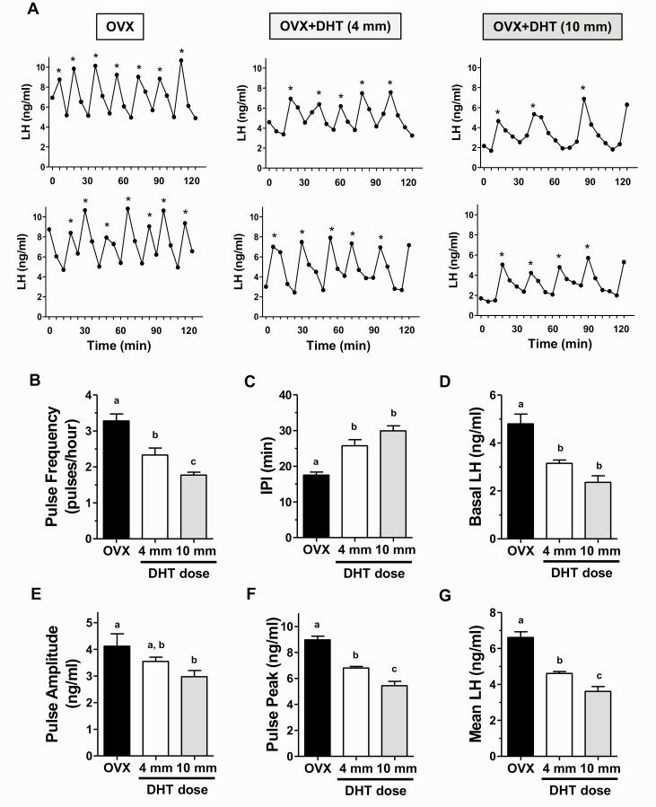
Figure 3.
DHT treatment reduces endogenous LH pulse secretion in young adult OVX females. (A) Representative profiles of in vivo LH secretion in DHT-treated OVX female mice (middle and right columns), and control OVX littermates (left column). LH was measured in serial tail-tip bleeds from awake, unrestrained females every 6 minutes for 2 hours. Identified pulses are indicated by *. Mean LH pulse frequency (pulses/hour) (B), interpulse interval (IPI; the number of minutes between pulses) (C), basal LH level (D), pulse amplitude (E), pulse peak (zenith value of a pulse) (F), and mean LH across the entire sampling period (G) were all significantly different in OVX+DHT vs control OVX females. Different letters above bars indicate significantly different (P < 0.05) from each other.

