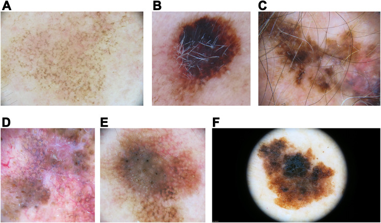
Figure 2.
Lentigo maligna(LM)/LM melanoma (LMM) dermoscopic findings. (A) Facial LM with an annular-granular pattern consisting of brown dots scattered around adnexal openings. (B) LM showing a large blotch with the blue-black sign. (C) LM on the scalp with follicle obliteration and central regression (gray peppering or granularity). (D) Facial LM with an annular-granular pattern around adnexal openings and follicle obliteration. (E) Brown lines coalescing to form rhomboids (angulated lines) around adnexal ostial openings at the periphery of this LM. Also, an asymmetric distribution of pigment surrounding the follicular openings can be seen. (F) Extrafacial LM with presence of network and atypical dots. Note the absence of adnexal openings in extrafacial skin.

