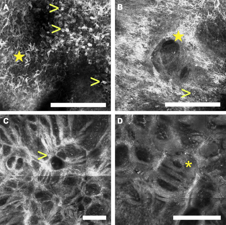
Figure 4.
Reflectance confocal microscopy images of a lentigo maligna. The epidermis (depth ~50 µm) shows numerous pleomorphic pagetoid cells composed of large round cells (arrowheads, A) as well as dendritic cells (star, A), which can invade the hair follicles (star, B). At the dermal-epidermal junction (depth ~150 µm), one can also identify disarrangement of the basal layer as well as large plump cells which correlate with melanophages (arrowhead, B), and junctional thickenings than can radiate from the hair follicles adopting a medusa head-like () structure (arrowhead, C), or a mitochondria-like structure (asterisk, D). (white scale bars: 250 µm).

