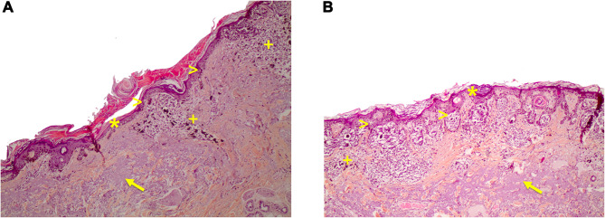
Figure 5.
(A) Histological pictures of lentigo maligna. Lentigo maligna in the radial growth phase. Solar elastosis (arrow), a patchy lymphocytic infiltrate and melanophages are seen in the dermis (+ sign). An atrophic epidermis (asterisk) and an increased density of melanocytes along the dermal-epidermal junction can be seen, some of them with variable nuclear atypia (arrowheads). Hematoxylin-eosin stain10x. (B) Extrafacial lentigo maligna on chronically sun-damaged skin. Atrophic epidermis (asterisk), prominent solar elastosis (arrow) and increased density of junctional pleomorphic melanocytes forming asymmetric nests with pagetoid growth of single melanocytes (arrowheads). Dermal inflammatory infiltrate with melanophages (+ sign). Hematoxylin-eosin stain10x.

