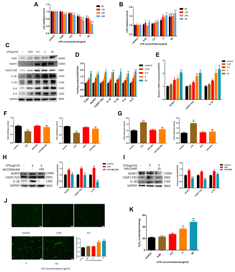
Figure 1.
LPS-induced NLRP3 inflammasome-dependent pyroptosis by the activation of ROS in HGFs. (A) Cell viability and (B) relative LDH release in HGFs treated by 0, 0.05, 0.5, 5 and 50 μg/mL of LPS for 6 h, 12 h, 24 h and 48 h. (C and D) The protein content in HGFs treated with 0, 0.05, 0.5, 5, 50 μg/mL of LPS for 6 h. (E) Relative mRNA expression was shown. (F) Cell viability and (G) relative LDH release in HGFs treated with 5 μg/mL LPS for 6 h with or without MCC950 (100 nM) or NAC (20 mM) for 1h.(Hand I) The protein content was shown. (J) Relative ROS and (K) H2O2 content in HGFs treat with 0, 0.05, 0.5, 5, 50 μg/mL of LPS for 6 h. Data were shown as mean ± SEM from three experiments independently. ***P < 0.001, **P < 0.01 and *P < 0.05 compared with control group. ###P < 0.001, ##P < 0.01 and #P < 0.05 compared with the group only treated by 5 μg/mL LPS.

