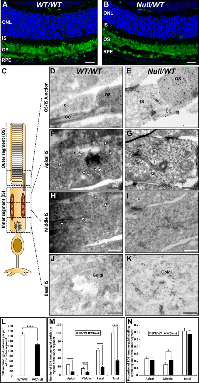
Fig 6. Reduced rhodopsin in the outer segment is not accompanied by accumulation in the inner segment.
(A, B) Rhodopsin staining (green) of 12μm retinal sections of 6-month-old heterozygous null female mice (Chmnull/WT) revealed strongly positive outer segments (OS), a small number of rhodopsin positive punctae (phagosomes) in the retinal pigment epithelium (RPE) but no detectable staining in the inner segment (IS). This was despite evident photoreceptor loss, as indicated by reduced numbers of DAPI stained (blue) nuclei in the outer nuclear layer (ONL). (C-K) Eye cup-derived specimens from 6-month-old heterozygous null female mice (Chmnull/WT) and age-matched controls were stained for rhodopsin by cryo-immunoEM using antibodies to the C-terminus (1D4-5nm gold) and N-terminus (RetP1-15nm gold). Rhodopsin was concentrated on the outer segment (OS) discs and the limiting membrane of the connecting cilium (CC). Rhodopsin labelling (examples indicated by white arrowheads in F-I) was also present on the Golgi and on small vesicles within the inner segment. Scale bars: 20 μm (A,B) and 200nm (D-K). (L,M) Quantitation of the number of 1D4 rhodopsin gold particles/area of IS and OS revealed that rhodopsin concentration was reduced to a similar extent in IS and OS of heterozygous null female mice (Chmnull/WT). (N) Dividing the IS into equal basal (containing the Golgi), middle and apical domains revealed a shift in the distribution of gold particles in the IS to a greater proportion in the middle region (mainly vesicle-associated). T-test: *p<0.05, ****p<0.0001, n = 3.

