Figure. 6.
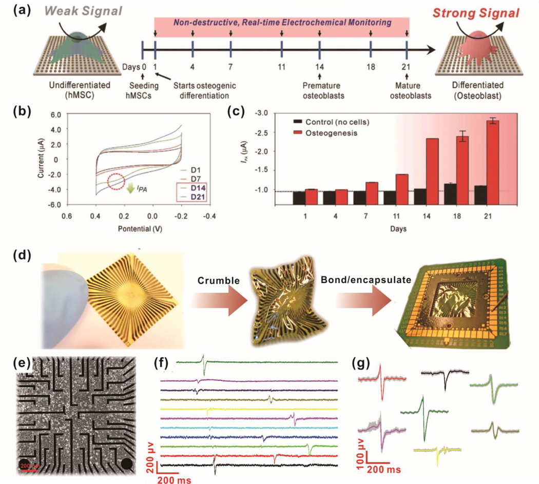
Non-destructive, live-cell monitoring methods using electrochemical sensing (a-e) and electrophysiological sensing (d-g). (a) Schematic diagram of electrochemical signal change during osteogenic differentiation of MSC on the nanoarray composed of gold and reduced graphene oxide. (b) Cyclic voltammogram of cultured MSC on the nanoarray from time-dependent monitoring (Day 0 to Day 21). (c) The cathodic peak currents of MSC cultured nanoarray from day 1 to day 21. (d) Tested graphene microelectrodes for heart tissue recording. A flexible chip was crumbled to mechanical deformation, then soldered and encapsulated. (e) Picture of HL-1 cells seeded on graphene microelectrodes. (f) Time trace recordings of HL-1 cells on 11 different channels of graphene microelectrodes. (g) The variety of recorded action potential shapes from different HL-1. (a)-(e) are reprinted with permission from ref. 28. © 2018 WILEY‐VCH Verlag GmbH & Co. KGaA, Weinheim, and (d)-(g) are reprinted with permission from ref. 101.

