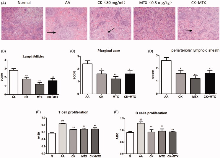
Figure 4.
Combined effects ofCK and MTX on spleen histopathology and proliferation of T cells and B cells. (A) Photomicrographs of the rat spleen (original magnification × 100, haematoxylin and eosin stain), (B) Lymphoid follicles, (C) Marginal zone, (D) the periarteriolar lymphoid sheaths, (E) T cell proliferation, (F) B cell proliferation. Data are expressed as the mean ± SD, with 6 animals in each group, ##p < 0.01 vs. Normal; *p < 0.05; **p < 0.01 vs. AA.

