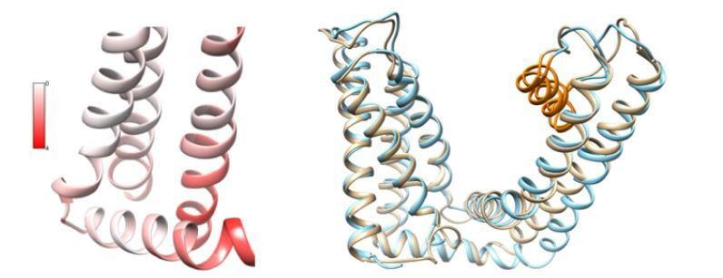
Author response image 1. Comparisons between 3J5R and 5IRX.

(Left) Per-residue structural deviation is plotted for the RMSD in Å between these two structures in the ligand-binding pocket. (Right) Structural overlay of 3J5R (cyan) and 5IRX (brown) between S1 and TRP helix.
