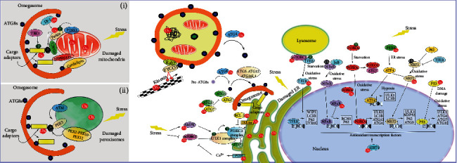
Figure 1.

Redox regulation of autophagy. Free radicals in the cell are mainly generated in mitochondria, peroxisomes, and ER; thus, a tightly regulated process to ensure proper functionality and turnover is crucial for cell survival (i.e., degradation by selective autophagy, e.g., mitophagy (i) or pexophagy (ii)). Under certain conditions (e.g., oxidative damage), autophagy is induced as an antioxidant pathway, and this leads to the initiation and nucleation of autophagy assembly sites (e.g., at the ER), with subsequent formation of the autophagosome, and eventual fusion with a lysosome to form a degradative autolysosome. ROS/RNS have the potential to regulate autophagy via upstream regulators, including proteins involved in the UPR system and the autophagy inhibitor mTOR, as well as redox modification in the cytoskeleton, affecting autophagosome transport. In addition, direct modifications in proteins involved in the autophagy process have also been identified including those involved in ATG8 cleavage and conjugation (i.e., ATG4 involved in LC3 cleavage; ATG3 and ATG7 involved in ATG8 lipidation), PI3KC3 activation and cargo recognition (e.g., p62/SQSTM1), and in selective autophagy (e.g., ATM in pexophagy and PINK1, Parkin and DJ-1 for mitophagy) (see the text for full description). Finally, autophagy and redoxtasis crosstalk is evident at the transcriptional level, with several transcription factors involved in autophagy regulation subject to redox modification. Some transcription factors regulate both redox levels and the autophagy process (e.g., NRF2, FOXOs, and p53). P (green): highlights phosphorylation events; Ub (black): highlights ubiquitination events; Ox (red): highlights sites for redox regulation of autophagy.
