Figure 2.
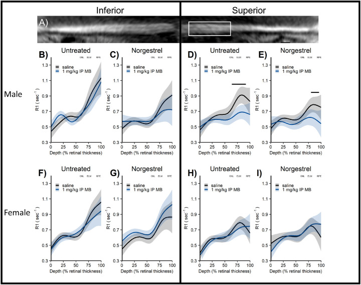
Central retina oxidative stress measured with QUEST MRI in Norgestrel-treated P23 dark-reared Pde6brd10 mice. (A) Representative flattened MRI image showing thinner superior retina in far periphery; white box indicates retina regions analyzed (± 400 – 1400 µm from ON although only superior region-of-interest shown for clarity). (B – E) Male P23 Pde6brd10 mice show in vivo oxidative stress limited to superior outer retina (untreated QUEST MRI); this finding is unmodified by Norgestrel (Norgestrel treated QUEST MRI) and there is little evidence for microglia activation (Iba-1 staining) in central retina. (F – I) Female P23 Pde6brd10 mice do not show evidence of oxidative stress (untreated QUEST MRI; Norgestrel treated QUEST MRI). To help spatially orient the MRI data, approximate location of outer nuclear layer (ONL), external limiting membrane (ELM), and retinal pigment epithelium (RPE) are noted based on representative OCT images from age, side, and sex appropriate untreated Pde6brd10 mice (OCT data not shown for clarity).

