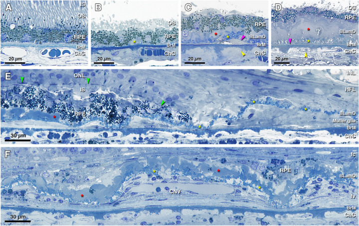
Figure 2.
Histopathology of basal laminar deposits (BLamD) in age-related macular degeneration (AMD). (A) Normal macula, without BLamD in a 77-year-old woman, 2000 µm nasal to the foveal center. (B) Patchy early BLamD (yellow asterisk) in a normal macula in an 88-year-old man, 600 µm nasal. (C) Early (yellow asterisk) and late (red asterisk) BLamD, along with basal linear deposit (fuchsia arrowhead), in an 85-year-old woman with early to intermediate AMD. Yellow arrowhead, ChC ghost, 400 µm temporal. (D) Thick, continuous early and late BLamD (yellow and red asterisks) in an 88-year-old man with early to intermediate AMD. A thin layer of pre-BLinD (fuchsia arrowhead) can be observed in this eye. Yellow arrowhead, ChC retraction, 290 µm nasal. (E) Persistent BLamD in the atrophic zone (yellow asterisks) of a 76-year-old woman with geographic atrophy. Müller glial processes were observed in the sub RPE-BL space. Note the continuity of BLamD across the border of atrophy, signified by the descent of the external limiting membrane (green arrowheads), 400 µm nasal. (F) Persistent BLamD overlying choroidal neovascularization (CNV) in a 91-year-old female. Both early and late (yellow and red asterisks) BLamD can be observed. Fibrovascular (fv) and fibrocellular (fc) membranes are shown on either side of the persistent BLamD, 490 µm temporal. INL, inner nuclear layer; HFL, Henle fiber layer; ONL, outer nuclear layer; IS, inner segment; OS, outer segment; RPE, retinal pigment epithelium; BrM, Bruch's membrane; ChC, choriocapillaris. Retina is detached from RPE in panels B to D.

