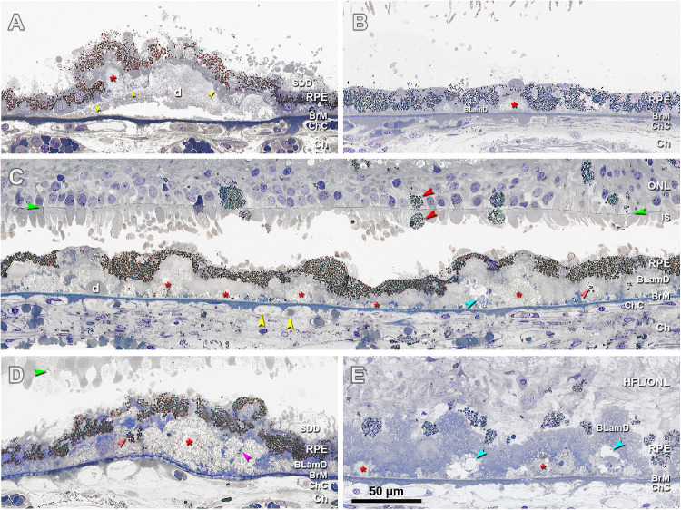
Figure 3.
Histopathology of basal mounds (BMounds) in age-related macular degeneration (AMD). (A) BMounds (red asterisk) are internal to the RPE basal lamina (yellow arrowhead) whereas drusen (d) are external to it, in a 76-year-old woman with early to intermediate AMD, 200 µm nasal. (B) BMounds (red asterisk) may appear as focal mound-like excrescences, in a 95-year-old man with early to intermediate AMD, 400 µm nasal. (C) BMounds (red asterisk) may form a semicontinuous layer of lipoprotein rich material within the BLamD. Pigmented cells of RPE origin can be observed in the retina and subretinal space (red arrowheads). Yellow arrowheads, retracted ChC and ghosts, in an 83-year-old woman with outer retinal atrophy, 1500 µm nasal. (D) BMounds can become quite large (red asterisk) and include complex features, such as internal structure (fuchsia arrowhead) and non-nucleated granule aggregates shed from RPE (orange arrowhead) in an 85-year-old woman with early to intermediate AMD, 1500 µm temporal. (E) Much like drusen, BMounds (red asterisk) may become calcified (blue arrowhead) in a 96-year-old woman with early to intermediate AMD, 2000 µm temporal. HFL, Henle fiber layer; ONL, outer nuclear layer; HFL/ONL, dyslamination of HFL and ONL; IS, inner segment; RPE, retinal pigment epithelium; BLamD, basal laminar deposit; BrM, Bruch's membrane; Ch, choroid; ChC, choriocapillaris. Green arrowhead, external limiting membrane. SDD, subretinal drusenoid deposits; Retina is detached from RPE in panels A to D.

