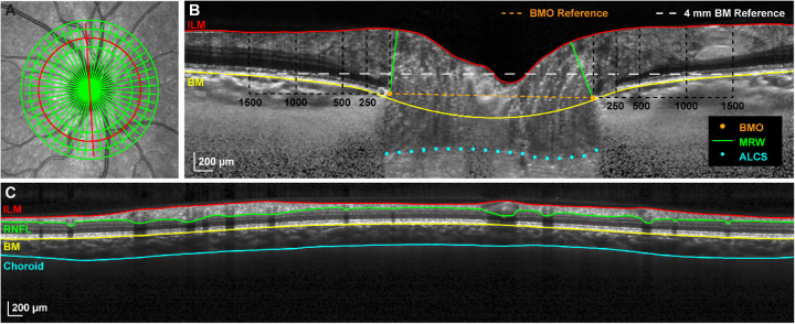
Figure 1.
Optical coherence tomography segmentation and parameters. (A) A 20° SLO image illustrating the custom composite OCT scan consisting of a 24-line radial scan and four circular scans (diameters 2.7, 3.5, 4.2, and 4.9 mm) centered on the ONH. (B) Compensated radial B-scan corresponding with the location of the red line in A. ILM and BM segmentations, manually selected BMO and ALCS points, BM and BMO reference planes, and locations of concentric annuli for TRT quantification (BMO to 250 µm, 250 to 500 µm, 500 to 1000 µm, and 1000 to 1500 µm) are shown. (C) Circular B-scan corresponding with the location of the 3.5 mm diameter red circle in A and illustrating ILM, RNFL, BM, and choroid segmentations.

