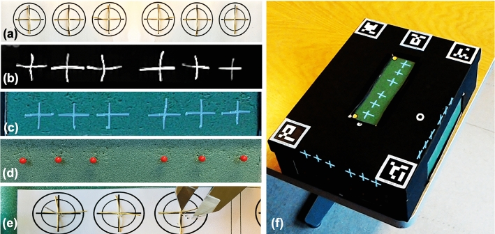
Fig. 2.
Overview of experimental workflow. a Anatomical internal targets. Wire crosses attached at the inner walls of the MRNOS. b Wire targets visible on CT scan dataset. c 3D model “Hologram” of corresponding wire crosses visible from the outside of the MRNOS. d Needle pins penetrated the 0.8 mm foam to meet the target with the guidance of the hologram. e View of the needle and the target from the inner wall of the MRNOS. Digital Caliper visible. f Hologram automatically registered/embedded into the MROS before the targeting task

