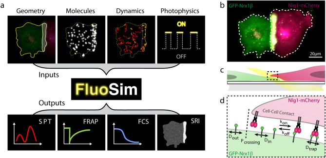
Figure 1.
Schematics of the simulator and experimental system. (a) General principle of FluoSim. (b) Contact between two COS cells, one expressing GFP-Nrx1β (green) and the other expressing Nlg1-mCherry (magenta), resulting in molecule accumulation through adhesive interactions (yellow zone). (c) Diagram showing a zoomed section of the cell–cell interface at the coverslip; the yellow beam represents oblique laser illumination. (d) Cartoon of Nrx1β diffusional trapping by Nlg1 in a cell–cell contact.

