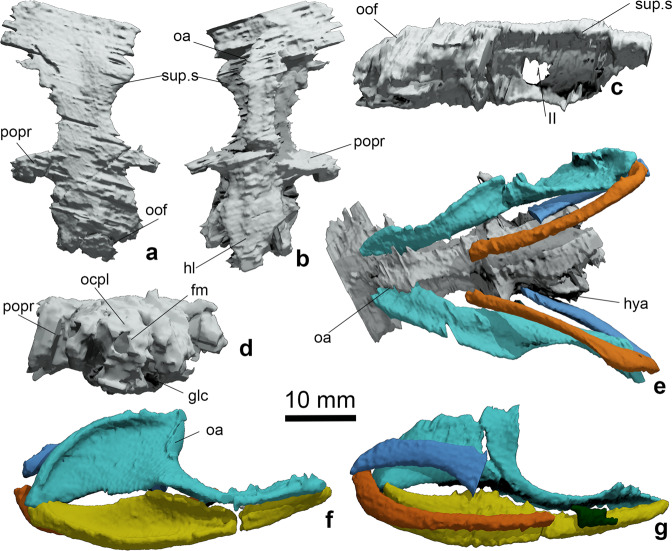
Fig. 3. Neurocranium and jaws of Ferromirum oukherbouchi gen. et sp. nov., PIMUZ A/I 4806.
Neurocranium in a dorsal, b ventral, c lateral, and d posterior, occipital views. e Articulation between braincase, palatoquadrate and hyoid arch in ventral view. f, g Arrangement of mandibular and hyoid arches in lateral and medial views respectively. Colour coding: grey, braincase; turquoise, palatoquadrate; yellow, Meckel’s cartilage; dark green, hypohyal; light blue, hyoid; orange, ceratohyal. Note that the rostral roof includes an excess of poorly resolved cartilage or matrix left in place in the computer renderings. fm, foramen magnum; hl, hypotic lamina; hya, hyomandibular articulation; glc, glossopharyngeal canal; oa, orbital articulation; ocpl, occipital plate; oof, otico-occipital fissure; popr, postorbital process; sup.s, supraorbital shelf; II, optic nerve.

